The importance of immunoassays in modern health care.
lmmunoassays are a cornerstone of contemporary medical diagnostics and research, offering precise, reliable, and versatile methods for detecting and quantifying a wide range of biological molecules. They derive their efficacy from leveraging the specificity of antibodies to bind target antigens. Generally speaking there are 3 current techniques with slight variations in applications namely, ELISpot, ELISA, and Flow Cytometry (including Fluorescence-Activated Cell Sorting or FACS) represent critical technologies within this domain, each having unique applications and contribution to healthcare.
What are they actually doing?
Immunoassays and Phenotyping
Phenotyping is the process of identifying cells by attributes or characteristics, some of these might be physical for example size and shape can vary significantly. An obvious example would be to compare a red blood cell with a muscle tissue cell where we know a red blood cell on maturity does not present a nucleus in favour of efficient oxygen exchange. Once differences go beyond physical we can use immunoassays to determine differences by specific biomarkers such a proteins. Our ability and approach to detecting different markers varies significantly, for example ELISpot and ELISA are relatively simple in terms of technical equipment requirements. They focus almost exclusively on secretion markers and in most cases are end-point assays requiring cells are washed away at some point within the process. Flow Cytometry and particularly FACS use a much more high tech approach in terms of equipment and mainly focus on markers that are presented on the surface of a cell. There are obvious cost implications to a process which requires highly specialised equipment however there are significant advantages to FACS such as the ability to sort cells and retrieve populations of cells with a specific target characteristic.
Enzyme-Linked ImmunoSpot or ELISpot
-
ELISpot is a highly sensitive immunoassay designed to detect secreted cytokines, antibodies, or other immune responses at the single-cell level. It involves capturing secreted molecules onto a solid surface, often using a specific capture antibody, then visualizing the spots where these molecules are present. Put most simply, cells are placed in a container and allowed to settle onto the surface of a membrane with a specific antibody for the target secretion molecule. Cells are then washed away leaving just the antibody bound secretion molecules on the membrane surface which can then be visualised by enzyme driven reaction. Whilst spot size can give an indication of levels of expression ELISpot is used for quantification of cells with the specific target expression.
-
Infectious Disease & Vaccines: ELISpot is extensively used to evaluate cellular immune responses to pathogens such as viruses and bacteria. Measuring T cell responses, for example, is crucial in understanding immunity and vaccine efficacy.
Cancer Research: It helps in assessing the activity of immune cells, including the effectiveness of immunotherapies that stimulate the patient's immune system to target cancer cells.
Autoimmune Diseases: ELISpot can detect autoreactive T cells, aiding in the diagnosis and study of autoimmune conditions.
-
Labour Intensive: An ELISpot assay can take multiple steps of labour intensive work and be technically demanding on the user requiring precision technique and timing.
Quantitative vs Qualitative: ELISpot specifically only gives quantitative data for the frequency of target molecule secreting cells and no specific data of high or low expression. Moreover in the main part because cells are often washed away in the process of developing the detected dots, often the result is an implied quantity rather than finite number.
Secretion Marker Specific: Reliance on binding of secretion proteins onto specific membrane bound antibodies means that cells surface markers can't normally be the target of the assay.
Enzyme-Linked Immunosorbent Assay or ELISA
-
ELISA is a versatile and widely used immunoassay that quantifies the presence of antigens (such as proteins, peptides, hormones, and antibodies) in biological samples. In most simple form it's a method to detect concentration of a target molecule in a solution. The method involves an enzyme conjugated to an antibody or antigen, which produces a measurable signal in response to the binding event. An important distinction between ELISpot and ELISA is that whilst ELISpot quantifies cells ELISA does not, however ELISA give quantifiable data of concentration of target expression (for example ELISA can detect differences in secretion between otherwise like for like samples before or after stimulation).
-
Diagnostic Applications: ELISA tests are standard for diagnosing infections like HIV, hepatitis, and Lyme disease, as well as monitoring hormone levels, allergens, and markers of inflammation.
Research: It facilitates understanding protein protein interactions, quantifying biomarkers, and conducting various biological and clinical studies.
Vaccine Development: ELISA assays are indispensable in measuring antibody responses and ensuring the efficacy and safety of vaccines.
-
Labour Intensive: Much like ELISpot assays there can be many labour intense and time dependant steps involved in carrying out ELISA assays.
Secretion Marker Specific: Mainstream ELISA are limited to secretion markers only.
Population Specific Data: ELISA is reliant on there being enough secretion marker present to elicit a response from and enzyme driven reaction to create a colour we can visualise. The assay can therefor only provide data on the expression of a marker from a population of cells not down to single cell.
Flow Cytometry & Flourescence-Activated Cell Sorting (FACS)
-
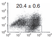
Flow Cytometry is a powerful technique for analysing the physical and chemical properties of cells or particles in a fluid as they pass through at least one laser. In simple terms cells are treated with a surface marker detection complex molecule then passed one by one through a laser detector which can detect the binding of the detector complex. FACS is a derivative technique that allows sorting of a heterogeneous mixture of cells into different populations based on specific light scattering and fluorescence characteristics. Cells are detected by multiple lasers and sorted into different populations by electrostatic deflection induced by the instrument according to positive or negative detection of the target marker.
-
Clinical Diagnostics: Flow Cytometry is crucial for diagnosing blood cancers like leukemias and lymphomas by identifying abnormal cell populations based on surface marker expression.
Immunophenotyping: It helps in characterising immune cells for research and clinical purposes, including monitoring HIV patient immune status by counting CD4+ T cells.
Therapeutics: FACS is used to isolate specific cell populations for therapeutic applications, such as stem cell transplantation and developing engineered T-cell therapies (e.g., CAR-T cells) for cancer treatment.
-
Labour Intensive: Requires sensitive sample preparation
Complex Equipment: A high reliance on expensive sophisticated equipment means a high barrier entry and technical expertise is required.
Data Complexity: Flow Cytometry and FACS produce complex graph data which requires complex interpretation or sophisticated software analysis.
Population Specific Data: Whilst multiple surface markers can be analysed data is population specific rather than individual cell and only populations of cells can be sorted rather than individual cells.
PhenoDot Addressing the Challenges
A primary focus of PhenoDot is keeping the system as simple as possible to avoid the high barrier to entry in training, maintenance, and cost of a system like FACS. Adopting this approach allows us to target the call of organisations like CEPI and the WHO with a system that is affordable and accessible for lower income countries and simpler laboratory setups. With this in mind we set about looking for ways to achieve more with systems like ELISA and ELISpot and there in lies our success with developing PhenoDot.
Base on the principals of ELISpot we have developed an assay in PhenoDot that allows cells to remain in the sample container (usually a 96 well plate but with PhenoDot this can be other platforms) and using our patent pending developing medium we can visualise cells according to the presence of either or both cell surface and cell secretion markers like no other immunoassay currently. In just 2 steps we’ve been able to overcome 2 of the biggest challenges in current immunoassay systems, firstly the high cost and barrier to entry. Reduced reliance on high tech equipment and as a result of better phenotyping reduced steps in cell identification so less overall assay required. Secondly we’re removing the detection limitations, as stated reducing the number of assays needed for positive identification of cells, improving workflows by use of a single assay type, and improved accuracy by detection on a single population of cells rather than parallel studies by different assays.
PhenoDot
Phenotyping like never before & unlocking new possibilities.
What this all means is with PhenoDot we’re able to do phenotyping like never before with a single assay which can identify according to both cell surface and secretion markers at the same time. We’re able to keep cells within the assay container, and with careful formulation of the developing medium maintain cell viability, allowing for single step identification and isolation of positively identified cells. Cells can simply be picked, washed removing the marker, and taken for the next step such as culturing for cloning or antibody generation. Whilst some of the labour intensive steps remain from all 3 alternative assays with PhenoDot we’re aiming to reduce the number of assays needed because we no longer need multiple assays to reliably identify cells by both surface and secretion markers.
With PhenoDot there’s opportunity to explore new possibilities of validating drug actions with assay that monitor the direct binding of the drug, the cell type by a surface marker and the cell response by a secretion marker. There are chances to develop new diagnostic systems based on disease states such as cancers with surface marker detection and potential to test cell response to chemotherapy with monitoring of secretions in response to the treatments.
Many of these latter mentioned assays are in development or still at the idea phase however the technology now exists making them a real possibility. The potential with PhenoDot are broad and vast which is why Elleon Biotech are looking for partnerships and collaborations to grow the many potentials into solid, robust assay kits.


